

Myeloma Australia
Services offered
Cancer information
Support from cancer nurses
Emotional support
+3
Tests for myeloma include blood tests, to measure your levels of paraprotein, and urine tests. You will also have imaging scans, such as CT scans, x-rays or an MRI, to check your bones.
You may have some genetic tests to look for gene faults in your blood or bone marrow. This information will help to guide your treatment.
You may also have a bone marrow aspiration, where a fluid sample is removed from the bone marrow with a thin needle, or a trephine biopsy, where a small piece of bone is removed. You will have a local anaesthetic and a mild sedative for these procedures.
You will usually have a full blood count along with other blood tests.
Serum analysis – There are two main blood tests that you are likely to have. Serum protein electrophoresis measures the level of paraprotein in your blood, while serum free light chain assay checks for a form of paraprotein known as free light chains.
Other blood tests – These check for low red blood cells (anaemia), too much calcium in the blood (hypercalcaemia), inflammation, and how well your liver and kidneys are working.
Once myeloma has been diagnosed, further blood tests will help your doctor work out the stage. Levels of paraprotein or free light chains can indicate changes in the activity of the myeloma. Your paraprotein and/or serum free light chain levels will be monitored to see how well treatment is working and to check that the myeloma is stable when you aren’t having treatment.
Blood tests are also an important way to check if the myeloma is changing your normal body function.
Myeloma cells produce an antibody known as paraprotein. To make a diagnosis of myeloma, doctors will look for this paraprotein in the blood or urine. Once you have been diagnosed with myeloma, doctors will regularly check your paraprotein levels to monitor the disease.
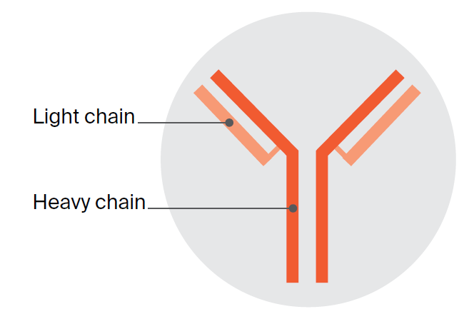
Paraprotein is made up of smaller proteins that are usually linked together to form a pair of long chains (called heavy chains) and a pair of shorter chains (called light chains). Most people with myeloma release complete (intact) paraprotein made up of both heavy and
light chains. Intact paraprotein can be detected in a blood test.
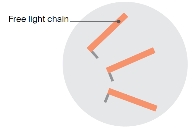
About 1 in 5 people with myeloma do not produce intact paraprotein. They produce only the light chain parts. These light chain parts are known as free light chains when detected in a special blood test, and as the Bence Jones protein when found in a urine test. Some people produce both intact paraprotein and free light chains.
Although uncommon, some people with myeloma produce little or no paraprotein. This is called non-secretory myeloma. It makes diagnosis and monitoring myeloma more difficult.
A urine test may be used to check for the Bence Jones protein, which is the light chain part of paraprotein. About one-third of people with myeloma make enough Bence Jones protein for it to show in the urine.
For this test, you may be asked to collect your urine in a large bottle over a 24-hour period. If the Bence Jones protein is detected, you may have regular urine tests to monitor the myeloma and check how well treatment is working.
Your doctor may also recommend that you have a specialised blood test called the serum free light chain assay (see Blood tests). This test may be done as well as, or instead of, a urine test, because the Bence Jones protein cannot be detected in a standard blood test.
These tests are used to examine cells from the bone marrow. They look for an increased number of plasma cells as well as abnormal plasma cells. You will usually have a local anaesthetic and a mild sedative.
For the bone marrow aspiration, a thin needle is used to remove a sample of fluid (aspirate) from the bone marrow – usually from the pelvic bone, but sometimes from the chest. A trephine biopsy, sometimes done with a second needle, removes a small piece of bone with the marrow inside.
The bone marrow samples are sent to a laboratory to be looked at under a microscope. A medical specialist, called a pathologist, will check the number of plasma cells in the sample and look for any damage to the bone marrow caused by myeloma.
Throughout treatment there may be times when bone marrow and trephine biopsies are repeated so that your doctors can see how well the treatment is working.
After a biopsy, you may feel drowsy and have some bleeding and discomfort at the biopsy site. It is a good idea to have someone drive you home after this test.
Your doctor will usually arrange imaging scans to check your bones. This may be done using a CT scan, x-rays or an MRI scan. Not all scans are available at all centres, and some scans may not be covered by Medicare. Speak to your doctor about any costs and availability.
A low-dose CT (computerised tomography) scan of the bones (also called a skeletal survey) is used to find any bone damage caused by the myeloma cells. For the CT scan, you lie flat on a table while the scanner takes detailed pictures of your body. The scan takes about 30 minutes.
Though less common, x-rays may be used instead of a CT scan. X-rays are usually taken of your head, spine, ribs, hips, legs and arms. Areas of bone damage, known as lytic lesions, show up as black shaded areas.
An MRI (magnetic resonance imaging) scan can find myeloma both inside and outside the bones. It uses magnetic fields and radio waves to take detailed cross-sectional pictures of the body. Let your doctor know if you have a pacemaker as the magnetic waves may interfere with it, though newer models do not have this problem.
For an MRI, you will lie on a table that slides into a large machine shaped like a cylinder. The scan may take 30–40 minutes. It is painless but can be noisy. You will be offered earplugs or headphones to listen to music. Some people feel anxious or claustrophobic in the cylinder. If you think you may feel distressed, let your medical team know before the appointment. You may be offered a mild sedative to help you relax.
CT and MRI scans of organs sometimes involve the injection of a dye (contrast) into a vein, usually in your arm. This dye makes the pictures clearer. Before having scans, tell the person doing the scan that you are suspected of having myeloma, as this dye can damage the kidneys and may not be safe for you. Also, let the medical team know if you have any allergies, if you have had a reaction to contrast during previous scans, if you have diabetes or if you could be pregnant. Skeletal surveys usually don’t need any contrast to be injected.
In some cases, your doctor may arrange specialised scans, such as a PET–CT (positron emission tomography–computed tomography) or sestamibi (MIBI) scan. Both of these scans involve an injection of a small amount of a harmless radioactive substance, which shows up areas of healthy and abnormal tissue. This may help detect myeloma outside of the bone marrow. Most people with myeloma do not need to have these scans.
Each cell in the body contains chromosomes, which are threadlike structures that hold sets of instructions known as genes. Every kind of cancer, including blood cancer, changes the genes of the affected cells. These gene faults are not the same thing as genes passed through families. The fault is only in the myeloma cells.
Genetic tests, such as fluorescence in situ hybridisation (FISH) and next generation sequencing analysis, look for abnormal chromosomes or genes in the blood or bone marrow.
There may be changes in the structure of the chromosomes within myeloma cells. Sometimes part of the chromosome breaks off or goes missing (deletion), or reattaches to another chromosome (translocation).
This information is used by your medical team to help guide treatment choices. Some specific chromosomal abnormalities are related to a higher-risk myeloma. Talk to your doctor about whether these tests are likely to be of use in your situation.
Myeloma Australia
Services offered
Leukaemia Foundation
Services offered
Find the right services for your needs, wherever you are in Australia
Trusted cancer information for all people affected by cancer
We're available 9am - 5pm, Monday to Friday (excluding public holidays)