

Breast Cancer Network Australia
Services offered
Cancer information
Emotional support
Physical symptoms and side effects
Tests to diagnose breast cancer include a physical examination, mammogram (breast x-ray), ultrasound and biopsy (taking a tissue sample).
The tests also show the size of the breast cancer and how far it has spread, which is called staging. Breast cancer can be stage 1, 2, 3 or 4. Stage 4 means that the cancer has spread to other parts of the body.
Extra tests on a biopsy sample may be done to look for certain genes, or other features (hormone receptors), which help plan treatment.
If you notice any breast changes or a swelling in your armpit, your GP will ask about your medical history and any family history of breast cancer. They will do a physical examination, checking both breasts and the lymph nodes in your armpit and above your collarbone. Your GP may also arrange some imaging tests, such as a diagnostic mammogram and/or an ultrasound and, if required, a biopsy. This is called a triple test.
Sometimes, a specialist will arrange these and additional tests, such as a breast MRI scan. You will also be referred for further tests if a screening mammogram has shown anything unusual.
Before a scan, tell the doctor if you have any allergies or had a reaction to dyes during previous scans. Also tell them if you have diabetes or kidney disease or are pregnant or breastfeeding.
A mammogram is a low-dose x-ray of the breast tissue. It can check any lump or other breast changes found during a physical examination. It can also show changes that are small or cannot be felt during a physical examination. If you have breast implants, it is important to let staff know before the mammogram.
Your breast is placed between 2 x-ray plates. The plates press together firmly to spread out the breast tissue so that clear pictures can be taken. You will feel some pressure, which can be uncomfortable, but the mammogram only takes 10–15 seconds. Both breasts will be checked.
Also known as three-dimensional mammography, tomosynthesis takes x-rays of the breast from many angles and combines them into a three-dimensional (3D) image. This may be better for finding small breast cancers, particularly in dense breast tissue.
This combines tomosynthesis with a dye (contrast) that is injected into a vein in your arm. A CEM may be helpful for people with dense breast tissue.
A national screening program provides a free mammogram for all women aged over 40. For more information, call 13 20 50 or visit BreastScreen Australia.
An ultrasound uses soundwaves to create a picture of breast tissue. It does not use radiation. It is often the first test done in women under 30 years with breast changes, or if a screening mammogram has picked up breast changes, or if you or your GP can feel a lump.
A gel will be spread on your breast, and then a small device (transducer) is moved over the breast and armpit. This sends soundwaves that echo when they meet something dense, like a tumour. A computer creates a picture from these echoes. The scan takes 15–20 minutes and is painless.
A magnetic resonance imaging (MRI) scan uses a large magnet and radio waves to take pictures of the breast tissue. It does not use radiation. It is mainly used for people at high risk of breast cancer or who have very dense breast tissue or breast implants. It may also be used if other imaging test results are unclear or to help plan surgery.
Before a breast MRI scan, you will usually have an injection of a dye (called contrast) to help show any abnormal breast tissue. You will lie face down on a table, which will slide into a large, cylinder-shaped machine. The scan can take up to 40 minutes. It is painless but loud, so you will wear earplugs. Some people feel claustrophobic. If you are concerned, talk to your doctor. You may be offered a mild sedative.
If breast cancer is suspected, a small sample of cells or tissue is taken from the lump or area of concern. A specialist doctor called a pathologist then checks the sample under a microscope for any cancer cells.
There are different ways of taking a biopsy and you may need more than one type. The biopsy may be done in a specialist’s rooms, at a radiology practice, in hospital or at a breast clinic. After any type of biopsy, your breast may feel sore and be bruised for a few days.
If tests on the biopsy sample confirm you have breast cancer, extra tests on the biopsy sample will be done to understand more about the breast cancer and help plan treatment. The results will be included in the pathology report.
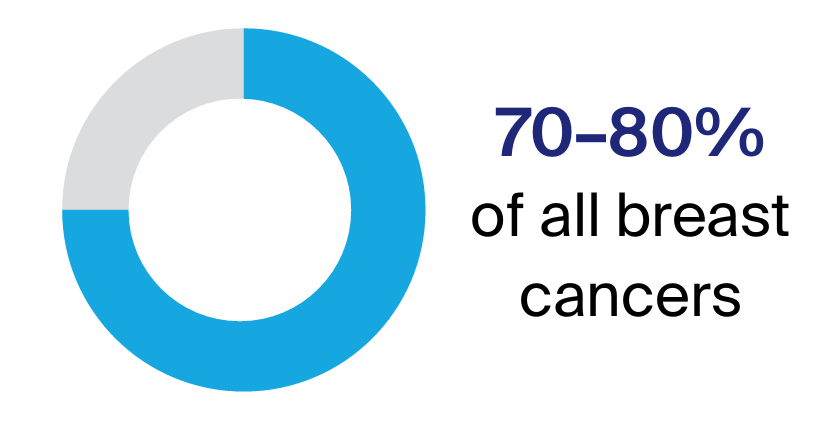
The hormones oestrogen and progesterone are produced naturally in the body. A receptor is a protein on the surface of the cell. Normal breast cells have oestrogen receptors (ER) and progesterone receptors (PR). Breast cancers that have these receptors are known as ER positive (ER+) or PR positive (PR+). This means that oestrogen or progesterone enters the cell, where it may stimulate cancer cells to grow. ER+ and PR+ cancers are usually treated with hormone therapy drugs (also known as endocrine therapy) that block the receptor, or drugs that reduce the amount of hormones that the body makes (aromatase inhibitors).
If the cancer has low levels of oestrogen receptors, hormone therapy drugs are sometimes used. These drugs are not used for cancers with no oestrogen receptors.
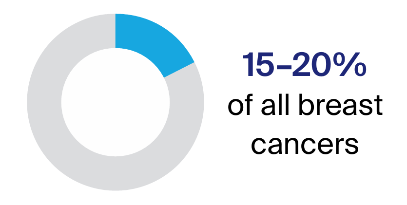
HER2 (human epidermal growth factor receptor 2) is a protein that is found on the surface of some cells and controls how cells grow and divide. HER2 levels are worked out with an initial test of the protein, and then can be confirmed with an in-situ hybridisation (ISH) test, which is done before giving targeted therapy. Tumours with high levels of these receptors are called HER2 positive (HER2+). Tumours with low levels are called HER2 negative (HER2– or HER2 low). It is often recommended that people with HER2+ breast cancer have chemotherapy and targeted therapy before they have surgery (neoadjuvant treatment). Depending on how the cancer responds to the neoadjuvant treatment and surgery, you may also have chemotherapy or targeted therapy after surgery (adjuvant treatment).
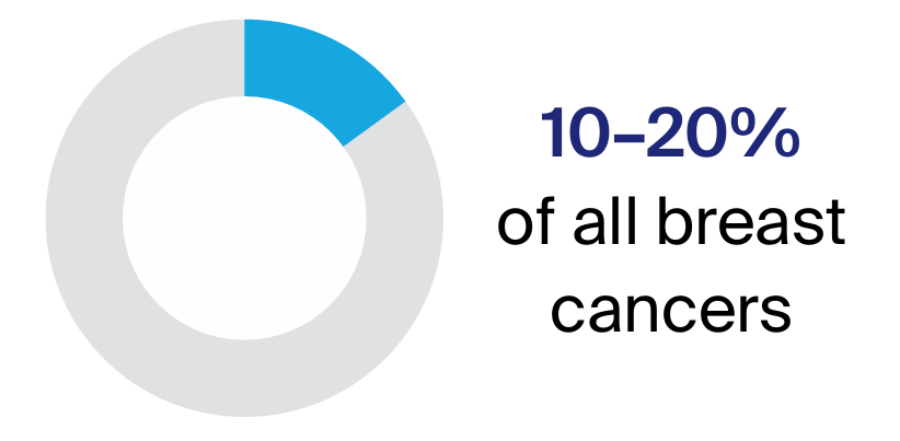
Some breast cancers do not have oestrogen (ER–), progesterone (PR–) or HER2 (HER2–) receptors. These are called triple negative breast cancers. Triple negative breast cancers do not respond to hormone therapy or to the targeted therapy drugs used for HER2+ cancers in early breast cancers. These types of cancer usually respond well to chemotherapy, so this may be used before and/or after surgery (neoadjuvant/adjuvant treatment). Some other types of targeted therapy may be used for triple negative breast cancer. Recently, various types of immunotherapy have been shown to work well for some triple negative cancers. These may be used before surgery for larger cancers or for cancers that also affect lymph nodes.
Gene expression profile tests may also be done on the biopsy sample, to help the doctors understand more about the cancer and the best way to treat it. These tests look at which genes are active in the cancer cells. The results provide information about the risk of cancer returning.
These tests may also be called genomic tests or molecular assays. They are different to genetic tests, which are used to look for inherited gene faults. For more information about genetic tests, see Does breast cancer run in families?
The gene expression profile tests available in Australia are Oncotype DX, EndoPredict, PAM50, and MammaPrint.
The test results will help the doctor work out if chemotherapy will be helpful after surgery. It can take 14 days for the results to come back, so it’s important to order these tests as soon as possible. It may be helpful for your surgeon to order these tests before you see your oncologist.
Ask your doctor if a gene expression profile test is an option for you. The standard pathology tests done on all breast cancers may be all that is needed for your treatment plan.
Gene expression profile tests may not be covered by Medicare; check what you may have to pay.
If tests show that you have breast cancer, you may have further tests to check whether the cancer has spread to other parts of your body. You will have a blood test to check your general health, and in some cases, it will test for specific tumour markers.
You may also have some of the following types of scans.
A bone scan is used to see if the breast cancer has spread to your bones. A small amount of radioactive solution is injected into a vein, usually in your arm. This solution is attracted to abnormal areas of the bone. After a few hours, the bones are viewed with a scanning machine. The scan is painless and the solution is not harmful.
A CT (computerised tomography) scan uses x-ray beams to take pictures of the inside of the body. Before the scan, dye (contrast) will be injected into a vein in your arm. This dye helps to make the pictures clearer. For the scan, you lie flat on a table while the scanner takes pictures. The scan takes about 30 minutes and is painless.
In a PET (positron emission tomography) scan, a small amount of low-level radioactive solution is injected into a vein in the arm or hand. Any cancerous areas take up more of the radioactive solution and may show up brighter in the scan.
Diagnostic tests can also show the size of the breast cancer and if it has spread to other parts of the body. This is called staging. It helps you and your health care team decide what treatment is best.
The most common staging system used for breast cancer is the TNM system. Letters and numbers describe how big the tumour is (T), if cancer has spread to nearby lymph nodes (N), or if it has spread to the bones or other organs, which is known as having metastasised (M).
The staging system also describes other details about the breast tumour such as oestrogen and progesterone receptor status, HER2 status and the grade of the cancer. Staging is usually done after surgery so the treatment team have full information about the cancer and whether it has spread to lymph nodes. The cancer may be classified as:
The grade describes how active the cancer cells are and how fast the cancer is likely to be growing.
Prognosis means the expected outcome of a disease. You may wish to discuss your prognosis with your doctor, but it is not possible for anyone to predict the exact course of the disease.
To work out your prognosis, your doctor will consider the stage and grade of the cancer, as well as features such as the cancer’s hormone receptor and HER2 status.
The survival rates for people with breast cancer have increased significantly over time due to more people taking part in breast screening, better tests and scans, and improved medicines and treatments. Doctors often use 5-year survival rates as a way to discuss prognosis. This is because research studies often follow people for 5 years; it does not mean you will survive for only 5 years. It also does not mean that the cancer cannot come back after 5 years.
Compared with other cancers, breast cancer has one of the highest 5-year survival rates when diagnosed early.
McGrath Breast Care Nurses are registered nurses specifically trained to support people with breast cancer from the time of diagnosis and throughout treatment. There are more than 200 McGrath nurses across Australia. For more information, visit McGrath Foundation.
Breast Cancer Network Australia
Services offered
McGrath Foundation
Services offered
Find the right services for your needs, wherever you are in Australia
Trusted cancer information for all people affected by cancer
We're available 9am - 5pm, Monday to Friday (excluding public holidays)