
Overview
Imaging scans use special equipment to create pictures of the inside of the body. They help doctors work out the stage of the cancer and the best treatment for you. Scans are done in a hospital or radiology clinic.

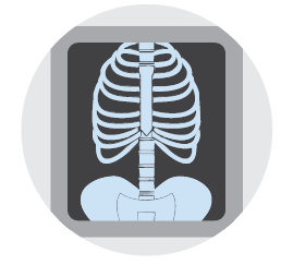
X-ray
An x-ray uses a small amount of radiation to create pictures of the inside of the body. The scan takes only a few minutes and does not cause pain.
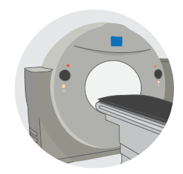
CT scan
A computerised tomography (CT) scan uses x-ray beams to take many pictures of the inside of your body. These images are combined into one detailed, cross-sectional picture. Before the scan, you may need to drink water or a special dye (called contrast). During the scan you may be injected with contrast. This makes the pictures clearer. You’ll need to remove any metallic objects, such as jewellery, before the scan. You’ll lie still on a table that moves in and out of a large, doughnut-shaped scanner. The scan takes only a few minutes. It’s painless but the contrast can cause a warm or flushing feeling.
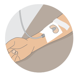
PET-CT scan
A positron emission tomography (PET) scan is sometimes combined with a CT scan to show the cancer in more detail. A low-dose radioactive glucose solution will be injected into your arm. You’ll then wait about 30–90 minutes for the solution to move through your body. You’ll need to remove any metallic objects, such as jewellery, before the scan. The scan takes 2–3 hours. A few hours after the scan, the solution will leave your body in your urine (wee). You may be told to avoid close contact with young children and pregnant women for a few hours.
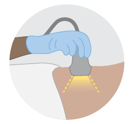
Ultrasound
An ultrasound uses soundwaves to create a picture of the part of the body that is affected by cancer. You’ll lie on a table and uncover the area being checked. A cool gel is spread on the skin, and a small handheld device (a transducer) is moved over your skin. Sometimes the transducer may be inserted into the body (e.g. to check the uterus). An ultrasound usually takes 10–20 minutes and doesn’t cause pain, but it may be uncomfortable. Before some ultrasounds, you may need to fast (not eat), or drink water to fill your bladder.
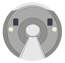
MRI scan
A magnetic resonance imaging (MRI) scan uses a magnet and radio waves to take detailed pictures of the body. Before the scan, you may be injected with a special dye (called contrast) to help show any cancer in the areas being scanned. Tell your doctor if you have any metallic implants (e.g. pacemaker), as some make MRI scans unsafe. You’ll also need to remove metal jewellery before the scan. You’ll lie very still on a table that slides into a tunnel-shaped scanner. The machine makes loud noises, so you’ll be given earplugs. The scan is painless and takes about 30–60 minutes.
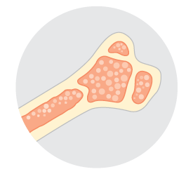
Bone scan
A bone scan helps show any abnormal areas of bone growth. You’ll be injected in the arm with a small amount of radioactive dye. You'll then wait for 2–3 hours while it moves through your bloodstream to your bones. Your body is then scanned. Areas of bone with cancer cells usually absorb more dye and show up more clearly. A bone scan can take up to 5 hours (including waiting time). The dye leaves your body in your urine (wee) after a few hours. You may be told to avoid close contact with children and pregnant women for the first few hours after the scan.
Before your scan, tell the doctor if you have any allergies or have had a reaction to dyes during previous scans. You should also let them know if you have diabetes, kidney disease, or if you are pregnant or breastfeeding.
Fear of small spaces
For CT, MRI and PET–CT scans, you’ll need to stay very still inside a large machine. If you feel anxious in small spaces (claustrophobia), let your cancer careteam know before your appointment. They can give you medicine to relax during the scan.
Find support services
Find the right services for your needs, wherever you are in Australia
Read cancer information
Trusted cancer information for all people affected by cancer
Need to talk to someone?
We're available 9am - 5pm, Monday to Friday (excluding public holidays)